
Protein A/G, cl-APC conjugate
$215.00
Cat. No: P02-P0117 Size: 100 ug
Protein A/G, cl-APC conjugated
Introduction:
Approximately 50.5 kDa. Recombinant Protein A/G consists of 7 IgG-binding domains EDABC-C2C3, which corresponds to Protein A and G domains that are included in the recombinant sequence. Protein A portion is from Staphylococcus aureus segments E, D, A, B and C. Protein G portion is from Streptococcus segments C2 and C3. The binding capacity of recombinant Protein A/G is broader than either Protein A or Protein G alone.
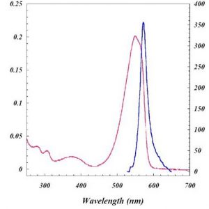
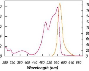
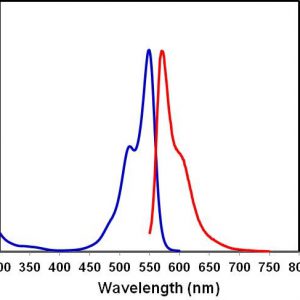
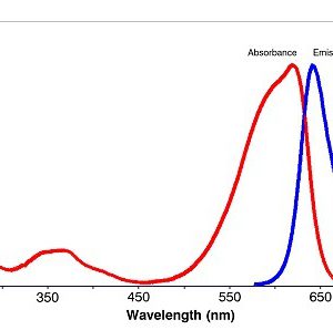
Allophycocyanin (APC), purified from Spirulina, is made up of alpha and beta subunits and is present as a trimer (αβ)3, which is unstable and susceptible to dissociation at low concentrations. The monomer, αβ, has a lower fluorescence quantum yield compared to the trimer and the maximal absorption is also shifted to 620 nm. The chemically cross-linked APC trimer (cl-APC) is much more stable than the native APC trimer, but still retains the same spectroscopic properties as the native APC trimer.
Protein A/G, cl-APC conjugated is a protein labeled by cl-APC.
Formulation: 100 µg of Protein A/G conjugated with cl-APC in PBS.
Excitation Laser: Red Laser (651 nm)
Application: Protein A/G is an excellent tool for purification and detection of mouse monclonal antibodies from IgG subclasses without interference from these other serum proteins. Individual subclasses of mouse monoclonals are most likely to have stronger affinity to this chimeric protein than to either Protein A or Protein G. Binding is less pH-dependent than either Protein A or Protein G alone, occurring well at pH 5-8.
Immunofluorescence (IF) 1: 50-1:200
Flow cytometry 1: 100-1:500.
The application notes include recommended starting dilutions; optimal dilutions/concentrations should be determined by the end user.
Usage:
This product is for research use only. It is not approved for use in humans, animals, or in vitro diagnostic procdures.
| Categories |
|---|
Be the first to review “Protein A/G, cl-APC conjugate”
Related products
Flow Cytometry reagents & Conjugates
Flow Cytometry reagents & Conjugates
Flow Cytometry reagents & Conjugates
Flow Cytometry reagents & Conjugates
Flow Cytometry reagents & Conjugates
Flow Cytometry reagents & Conjugates
Cell-based immunoassay kits

Flow Cytometry reagents & Conjugates
Reviews
There are no reviews yet.